When a weakened vertebra collapses, it can cause sudden back pain that flares every time you stand or move. This page explains what a compression fracture is, lets you step through how bone cement stabilizes and can re-lift the bone, and walks through the options — from bracing to kyphoplasty to fixation — plus how to protect against the next fracture.

A collapsed vertebra tips the spine forward
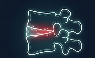
A break in which the front of a spinal bone collapses, most often because the bone has been weakened by osteoporosis. It is one of the most common fractures in older adults.
Each vertebra has a block-shaped front portion, the vertebral body, that carries most of your weight. When the bone inside becomes thin and porous from osteoporosis, that block can crack and collapse under everyday loads — bending, lifting a bag of groceries, or even a hard cough. Because the front of the body usually gives way first, it collapses into a wedge shape, which tips the spine a little further forward with each fracture.
The classic story is sudden, sharp back pain at a specific spot that is much worse with standing, walking, or changing position and better lying flat. Some fractures, though, cause surprisingly little pain and are found by chance on an X-ray. Unlike a herniated disc, a compression fracture usually does not pinch a nerve, so pain that travels down a leg, or new numbness or weakness, is a signal to look for a different or more serious problem.
Most compression fractures heal on their own over six to twelve weeks. The two questions that matter most are: does this painful fracture need a procedure to settle it, and — just as important — why did the bone break, and how do we prevent the next one? Both are covered on this page.
Osteoporosis thins the vertebral body until it cracks under ordinary loads, sometimes with no real injury.
A sharp pain at one spot that worsens with standing or moving and eases lying down is the classic sign.
Most fractures settle over 6 to 12 weeks with pain control and gentle activity, no procedure required.
One fragility fracture sharply raises the odds of another, so treating the underlying bone is essential.
A compression fracture usually does not affect the nerves. Seek prompt evaluation for new leg numbness or weakness, loss of bladder or bowel control, fever, unexplained weight loss, or a fracture after high-energy trauma. These can point to a burst fracture pressing on the spinal canal, or to a fracture caused by infection or a tumor rather than osteoporosis — situations that are managed very differently.
Cement augmentation is very good at the mechanical pain of a fresh fracture and less useful for old pain or established deformity. Select a symptom to see what to realistically expect.
Select a symptom to see how treatment typically affects it.
Pick a fracture pattern, then step through a vertebroplasty or a balloon kyphoplasty to see how bone cement stabilizes the vertebra — and why only kyphoplasty can restore some lost height. This is educational, not a diagnosis.
Whether a procedure helps depends on the fracture, its pattern, your bone health, and your general health. Choose a category, then select a factor to learn more.
Select a factor to see how it affects candidacy and which treatment fits.
Most fresh fractures are managed without a procedure. Cement augmentation is considered for pain that is not settling, and fixation is reserved for unstable fractures. Alongside all of these, the underlying bone is treated.
Most osteoporotic compression fractures heal on their own over six to twelve weeks. Short-term pain medication, a brace for comfort, and staying gently active while avoiding prolonged bed rest allow the bone to knit. At the same time, we start looking into why the bone broke.
When pain is severe or not improving with a fair trial of non-surgical care, injecting bone cement (vertebroplasty or kyphoplasty) can splint the fracture and relieve pain. Unstable or burst fractures, or those threatening the nerves, instead need screws and rods.
Select an option to see how it works and when it is chosen. At Brown we favor the least-invasive effective treatment and reserve bigger operations for genuine instability.
When a fracture needs more than cement, screws and rods stabilize the spine while it heals. See how those screws are placed through small incisions, using robotic navigation.
This walks through a typical balloon kyphoplasty — lifting the collapsed bone and filling it with cement. Select an option above to switch pathways, and select each step to learn what happens and why.
For the right fracture, cement augmentation is one of the more gratifying procedures in spine care. Results vary, and honest expectations matter.
Cement augmentation is generally low-risk, but no procedure is risk-free. The main specific concern is cement leaking outside the vertebra; usually this causes no problem, but rarely it can irritate a nerve or, very uncommonly, travel in the bloodstream. Other risks include infection, bleeding, and, importantly, a new fracture at a neighboring level — the bone next to a treated vertebra remains osteoporotic and still carries load. This is one more reason that treating the underlying bone is not optional.
It is also worth being clear about benefit: augmentation reliably targets mechanical fracture pain, but it does not treat pain from arthritis, muscle strain, or old healed fractures, and it does not reverse a stooped posture that has already set in. Careful selection — the right fracture, the right pain pattern, confirmed on imaging — is what separates a gratifying result from a disappointing one.
A compression fracture is often the first visible sign of osteoporosis. Fixing the fracture without treating the bone leaves the real problem in place — and if a fusion is ever needed, the strength of that bone is what the hardware has to hold onto.
Doctors call a fracture that happens from a minor force a fragility fracture, and it is a sentinel event: it tells us the bone is weak enough to break again. The risk of a second fracture is highest in the first year, and each new fracture makes the next one more likely and the spine a little more stooped. That cascade is preventable, and breaking it is as important as treating the fracture that brought you in.
For that reason, part of caring for a compression fracture is a bone-health work-up and, in most cases, medication to rebuild bone strength. This is usually coordinated with your primary care doctor or an endocrinologist, and it is the step most likely to protect your independence over the years ahead. It matters even more if spine surgery with screws (a fusion) is on the table, because weak bone is the single biggest reason hardware loosens and a fusion fails to heal.
A DEXA scan measures bone density and gives a T-score. If you have already had a recent CT of the chest, abdomen, or spine, the density of the bone on that scan (its Hounsfield units) can be read at no extra cost or radiation, and is often more reliable than DEXA when the spine has a lot of arthritis.
Adequate calcium and vitamin D, protein, strength and balance exercise, stopping smoking, and limiting alcohol all support bone and improve the odds that any surgery heals.
Blood tests look for treatable contributors to bone loss; when a fracture looks unusual, imaging helps exclude a tumor or infection.
Depending on your risk, a bone-building (anabolic) or bone-protecting (antiresorptive) medication is started. For higher-risk bone — and before a planned fusion — a bone-building drug started first is often the better choice.
Home-safety changes, vision checks, and reviewing medicines that cause dizziness reduce the falls that trigger fractures.
Which medication fits depends on your fracture risk, kidney function, dental health, cardiovascular history, and other factors, decided with your bone-health physician. This is general education, not a prescription.
These are the specific thresholds clinicians use to decide when bone is weak enough to act on, and when it is weak enough to change a surgical plan. They are shared here for transparency; your own targets are set by your physicians.
A fragility fracture — like a compression fracture — counts as osteoporosis on its own, whatever the T-score says. DEXA can also read falsely high when the spine has arthritis or calcified vessels, so a “normal” DEXA does not fully clear weak bone.
Measured from a CT you may already have. For spine surgery with screws, the risk of loose hardware and a fusion not healing climbs as this number falls; many surgeons treat roughly < 120 HU at the levels being fixed as a red flag worth optimizing before elective surgery.
Any one of these tips bone from “monitor” to “treat, and optimize before elective surgery”:
Most compression fractures never need screws. But when a fusion is planned in osteoporotic bone, treating the bone before the operation measurably improves the odds that it heals. Screws hold better in denser bone, cages are less likely to sink, and the fracture rate at the top of a construct falls.
The strongest evidence is for anabolic (bone-building) therapy. When the timeline allows, starting an anabolic such as teriparatide roughly 2–3 months before an elective fusion and continuing it for 6–12 months after has been associated with higher fusion rates (about 82% versus 68% with a bisphosphonate in one representative series) and fewer loose screws. The bone-building course is then followed by an antiresorptive to hold the gains.
This is always balanced against how urgent the surgery is: an unstable fracture or a nerve at risk is treated promptly and the bone is optimized around the operation rather than delaying it. The plan is made jointly by your spine surgeon and a bone-health physician.
Both inject bone cement to splint a painful fracture through a needle in the pedicle. The difference is whether a balloon is used first to make a cavity and lift the bone. The best choice depends on your fracture and is decided with your surgeon.
| Feature | Vertebroplasty | Balloon Kyphoplasty |
|---|---|---|
| What it does | Injects cement directly into the fractured bone | Inflates a balloon to make a cavity, then fills it with cement |
| Restores height? | Minimal | Can restore some lost height |
| Adds hardware? | No — cement only | No — cement only |
| Anesthesia | Local with sedation, or general | Local with sedation, or general |
| Cement leak risk | Slightly higher (no contained cavity) | Lower (cement placed into a cavity) |
| Consideration | Vertebroplasty | Kyphoplasty |
|---|---|---|
| Painful fracture, little height loss | Well suited | Also suitable |
| Painful fracture with height loss | Stabilizes but will not lift | Preferred; can re-lift the bone |
| Very collapsed / hard to reach | Sometimes chosen | Cavity creation can help |
| Unstable or burst fracture | Not appropriate alone | Not appropriate alone |
| Factor | Vertebroplasty | Kyphoplasty |
|---|---|---|
| Hospital stay | Usually same day | Usually same day |
| Pain relief timing | Often within a day or two | Often within a day or two |
| Posture correction | None expected | Some, if done early |
| Next-level fracture risk | Present; treat the bone | Present; treat the bone |
Neither is universally better. For a painful fracture with meaningful height loss we often favor kyphoplasty; for a painful fracture with little collapse, either can work well. Both depend entirely on choosing the right fracture in the first place.
This educational resource was developed by the Division of Spine and Spine Tumor Neurosurgery at Brown University Health to help patients and families understand vertebral compression fractures and their treatment. Our surgeons offer the full spectrum of care, from cement augmentation to stabilization for unstable fractures, and work closely with primary care, endocrinology, and physiatry so that both the fracture and the underlying bone are treated.


For full faculty profiles, visit the Brown Neurosurgery Spinal Surgery Division.