Spondylolisthesis is the forward slipping of one vertebra over the one below it. Depending on how far it slips and why, it can cause back pain, sciatica, or the leg-heaviness of a narrowed canal. Explore how the slip is graded, see how it pinches the nerves, and learn when it is watched, decompressed, or fused.
One vertebra slips forward over the bone below
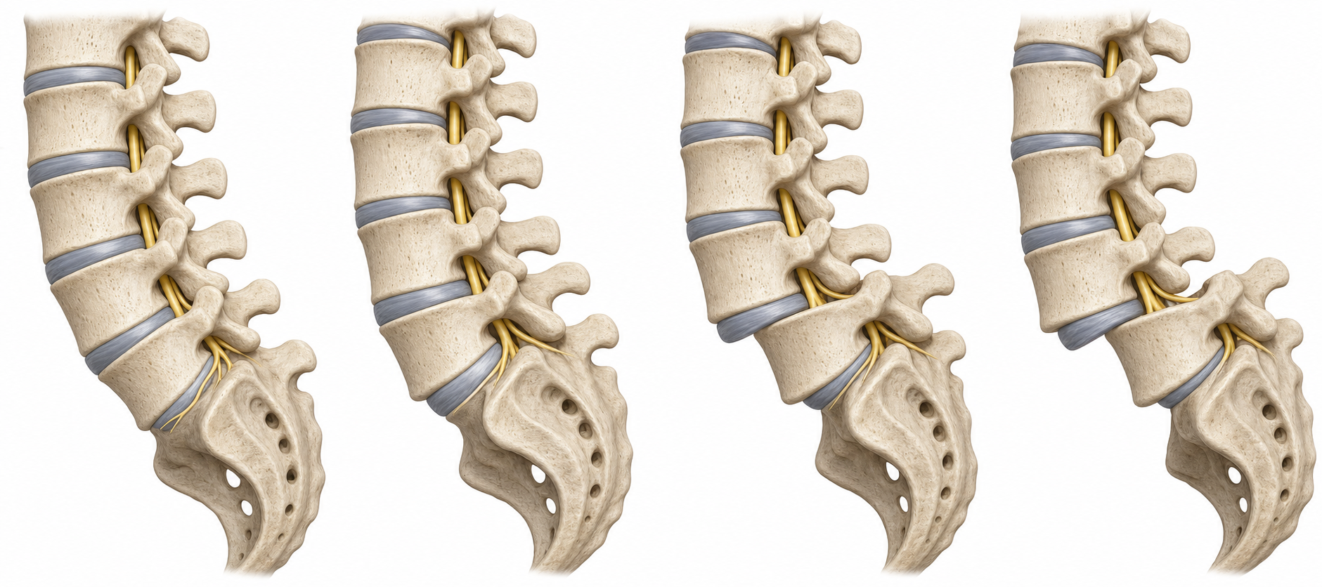
A condition in which one vertebra slides forward over the vertebra beneath it. How much it slips is described by a grade, and why it slips defines the type.
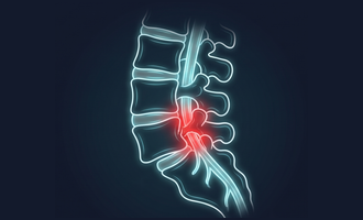
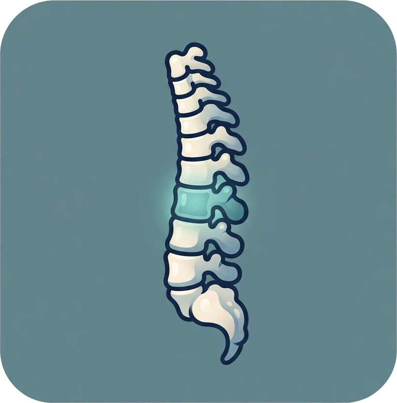
The spine is a stack of bones (vertebrae) held in line by discs in front and paired facet joints behind. In spondylolisthesis, one vertebra loses part of that restraint and slides forward on the one below. The slip may be tiny and silent, or large enough to crowd the nerves and cause symptoms.
Two types cause most cases. In isthmic spondylolisthesis, a small stress fracture in a bony bridge called the pars (a pars defect) lets the vertebral body slide forward while the back part of the bone is left behind; this most often happens at L5–S1 and can begin in adolescence. In degenerative spondylolisthesis, the disc and facet joints simply wear out with age and let the whole vertebra drift forward, most often at L4–L5; because the back of the bone moves too, it tends to narrow the central canal and cause the leg-heaviness of spinal stenosis.
The amount of slip is graded with the Meyerding scale from I to IV (roughly, each grade is another 25% of forward slip). Grade, type, symptoms, and whether the segment is stable or moves abnormally all shape treatment. At Brown, our approach is least-invasive-first: relieve the nerves with the smallest effective operation, and add a fusion when the segment is genuinely unstable. The interactive tool below lets you see how the slip and its type change what the nerves feel.
A stress fracture in a bony bridge lets the body slip forward while the back of the bone stays put, pinching the exiting nerve.
Aging discs and facet joints let the whole vertebra drift, narrowing the central canal like spinal stenosis.
The slip is graded I to IV by how far the vertebra has moved forward. Higher grades are less common.
Whether the segment moves abnormally on flexion/extension X-rays is the single biggest factor in deciding on fusion.
Most spondylolisthesis is not an emergency, but seek care right away for new loss of bladder or bowel control, numbness in the groin or inner thighs (the "saddle" area), or rapidly worsening leg weakness. These can signal cauda equina syndrome, a surgical emergency.
Spondylolisthesis is graded by how far the upper vertebra has slipped forward over the one below. Higher grades are less common and more likely to need surgery.
Illustration for education. Grade is one input into treatment; symptoms and stability usually matter more than the number alone.
Drag the slider to increase the forward slip, and switch the type, to see how the vertebra moves, how the grade changes, and where the nerves get pinched. This is educational only, not a measurement of your own spine.
This is a simplified illustration of how a slip crowds the nerves, not a measurement or diagnosis of your own spine.
Surgery is very good at the leg symptoms of pinched nerves and, when a segment is unstable, at the pain of that instability. Select a symptom to see what to realistically expect.
Select a symptom to see how treatment typically affects it.
Grade, type, stability, symptoms, and health all shape whether surgery makes sense and whether a fusion is needed. Choose a category, then select a factor.
Select a factor to see how it affects candidacy and whether fusion is needed.
Many people do well without surgery. When surgery is needed, the two questions are the same as always: do the nerves need more room, and does the segment need to be held together?
A low-grade, stable slip often responds to physical therapy focused on core and abdominal strength, activity changes, anti-inflammatory or nerve-pain medication, and, in selected cases, an epidural steroid injection. Many slips do not progress, so patience and rehabilitation come first.
Surgery is considered when leg symptoms are severe or persistent, when there is significant weakness, or when the segment is unstable. The nerves are decompressed; a fusion is added when the slip is unstable, higher-grade, or when decompression alone would risk further slipping.
Select an option to see how it works and when it is chosen. At Brown we favor the least-invasive effective approach and reserve fusion for genuine instability or deformity.
When a slip is fused, screws and rods hold the level in place while it heals. See how those screws are placed through small incisions, using robotic navigation.
This walks through a typical decompression with fusion (for example a TLIF) for an unstable slip. Select each step to learn what happens and why.
Surgery for symptomatic spondylolisthesis is one of the more reliable operations in spine surgery, especially for leg symptoms. Results vary, and honest expectations matter.
Every spine operation carries risk. For decompression, the most common specific risk is a small tear in the lining around the nerves (a dural tear), usually repaired during surgery. Other risks include infection, bleeding, nerve irritation, and, less commonly, new or persistent weakness or numbness.
Fusion adds its own considerations: a longer recovery, a small chance the bones do not fully knit (nonunion), which smoking raises, and adjacent segment disease, where the levels next to a fusion carry more load over the years and may wear faster. This is precisely why a stable, low-grade slip is often better served by decompression alone (or no surgery) rather than a fusion it does not need.
For a slip, this is the central decision. A stable slip and an unstable one are different problems that favor different answers, decided with your surgeon.
| Feature | Decompression Alone | Decompression + Fusion |
|---|---|---|
| What it does | Reopens the canal / foramen to free the nerves | Frees the nerves and joins the segment with a cage and screws |
| Adds hardware? | No | Yes: interbody cage plus pedicle screws |
| Preserves motion? | Yes, at that segment | No; the slipped segment is made solid |
| Addresses instability? | No | Yes; stops the abnormal movement and can partly reduce the slip |
| Best for | Stable, low-grade slip with leg-dominant symptoms | Unstable, higher-grade, or mechanically painful slips |
| Consideration | Decompression Alone | + Fusion |
|---|---|---|
| Stable segment | Often sufficient | Usually unnecessary |
| Unstable on flexion/extension X-rays | Risk of further slip | Preferred; stabilizes the segment |
| Mechanical back pain from the slip | Not addressed | Directly addressed |
| Higher-grade slip / deformity | Rarely enough alone | Preferred |
| Brown's default preference | Favored when stable | Reserved for genuine instability |
| Factor | Decompression Alone | + Fusion |
|---|---|---|
| Hospital stay | Often same day to one night | Typically 1–3 nights |
| Return to light activity | 1–2 weeks | Several weeks, with restrictions |
| Full recovery | A few weeks | 3–6 months as bone fuses |
| Long-term consideration | Possible progression of the slip | Adjacent-segment wear over years |
The aim is to match the operation to the slip: reopen the nerves with the least disruption, and add stability only when the segment truly needs it.
A fusion only succeeds if the bone heals around the hardware and keeps its grip on the screws. When bone is thin, screws loosen, cages settle into the vertebra, and the fusion can fail to knit — so when a fusion is part of the plan, strengthening the bone beforehand is part of the operation, not an afterthought.
Spinal hardware is only as strong as the bone it anchors into. In osteoporotic bone, pedicle screws pull out or loosen more often, interbody cages can sink into the soft vertebral body (subsidence), and the fusion is slower and less certain to form a solid bridge — a higher risk of pseudarthrosis, or non-union. Longer, multi-level constructs carry the most bone-related risk, including new fractures at the top of the construct. Weak bone rarely rules surgery out, but it changes how we plan and prepare for it.
Before an elective fusion we take stock of the bone. A DEXA scan gives a T-score, and a CT you may already have gives a direct read of bone density — in Hounsfield units — at the very levels we plan to instrument, which is often more reliable than DEXA when the spine is arthritic. Bloodwork checks vitamin D and screens for reversible causes of bone loss.
When the numbers are low and the operation is elective, we treat the bone first. The strongest evidence is for anabolic (bone-building) agents — teriparatide, abaloparatide, or romosozumab. When the timeline allows, starting an anabolic roughly 2–3 months before surgery and continuing it 6–12 months afterward is associated with higher fusion rates and fewer loose screws, and it is then followed by an antiresorptive to hold the gains. Building first and protecting second is the sequence that adds the most strength. Urgent or unstable cases are not delayed — there we optimize around the operation instead of before it.
Coordinated with your surgeon and a bone-health physician, and weighed against how urgent surgery is. General education, not a prescription.
The thresholds below are what teams use to decide when bone is weak enough to treat, and weak enough to optimize before an elective fusion. Shared here for transparency; your own targets are set by your physicians.
A prior fragility fracture counts as osteoporosis on its own. DEXA can read falsely high over spinal arthritis, so a normal DEXA does not fully clear weak bone before a fusion.
Read from a CT you may already have. Loose hardware, cage subsidence, and non-union climb as this falls; many surgeons flag roughly < 120 HU at the instrumented levels as worth optimizing before elective surgery.
Any one of these shifts an elective fusion toward treating the bone beforehand:
This educational resource was developed by the Division of Spine and Spine Tumor Neurosurgery at Brown University Health to help patients and families understand spondylolisthesis and its treatment options. Our surgeons offer the full spectrum of care, from minimally invasive decompression to fusion and deformity correction, and work closely with physical therapy, pain management, and physiatry so that surgery is recommended only when it is the right next step.


For full faculty profiles, visit the Brown Neurosurgery Spinal Surgery Division.