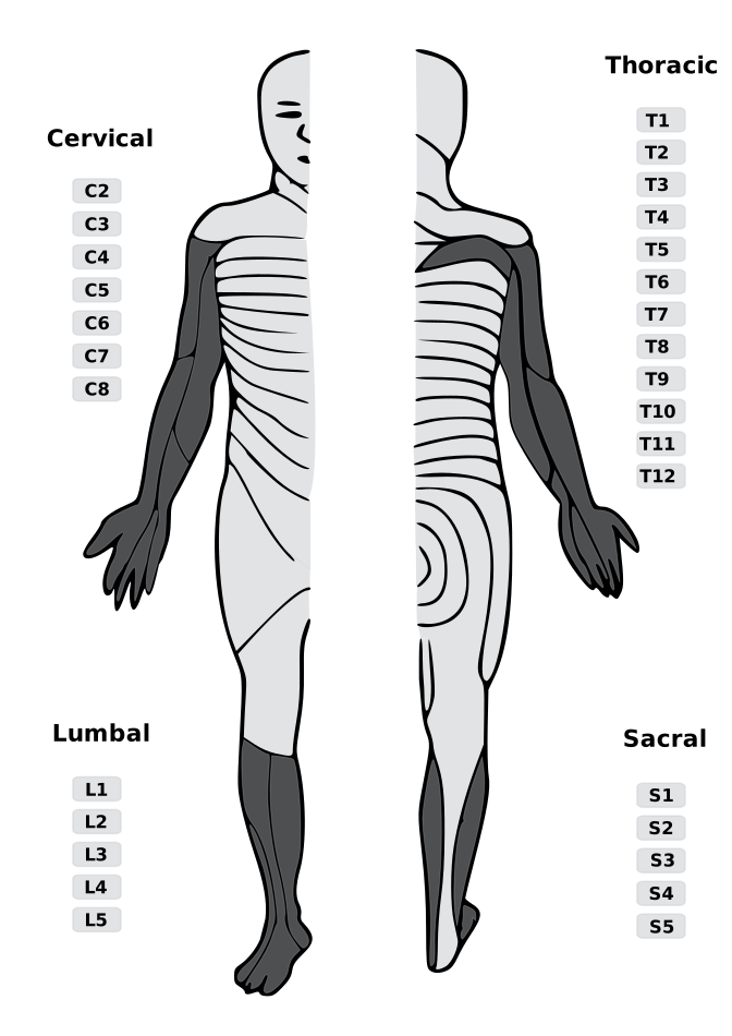
Lumbar/Sacral · Lower-extremity pattern aid
Lumbar disc level → nerve root deficit.
Select a disc level and herniation morphology. The tool returns the most likely affected root with its dermatome, myotome, reflex, and bedside clue.
Approximately 95% of lumbar disc herniations occur at L4–L5 or L5–S1 [StatPearls].
L4–L5
Dermatome (sensory)
Myotome (motor)
Reflex
Bedside clue
Lumbosacral sensory zones
Morphology atlas · Axial schematic
Disc shape changes which root is at risk.
Tap a morphology to morph the axial view and see where the fragment sits relative to the canal, lateral recess, and neural foramen. These are teaching schematics, not diagnostic MRI criteria.
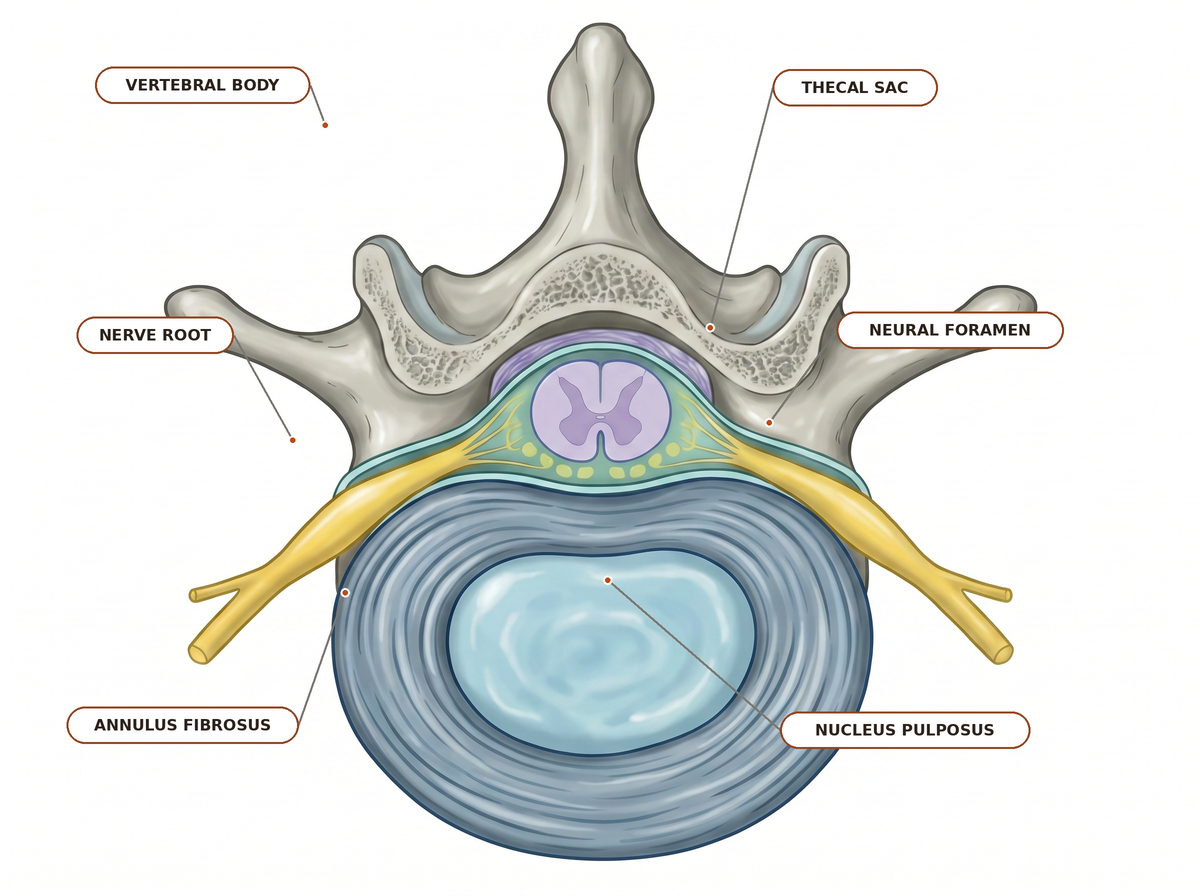
Normal axial relationship
Baseline before herniation: the posterior disc margin stays behind the thecal sac, with clear lateral recess and foraminal corridors.
Reference anatomy — the only labeled diagram.Traversing-versus-exiting root rules follow StatPearls — Lumbar Disc Herniation.
Comparison matrix
All five disc levels, both morphologies. Tap a row to load it in the tool above.
| Disc level | Paracentral → traversing root | Far-lateral → exiting root | Dermatome (autonomous zone) | Motor | Reflex |
|---|
Autonomous zones from StatPearls — Radicular Back Pain. Motor and reflex columns synthesize StatPearls — Lumbar Disc Herniation and the Merck Manual.